Slides
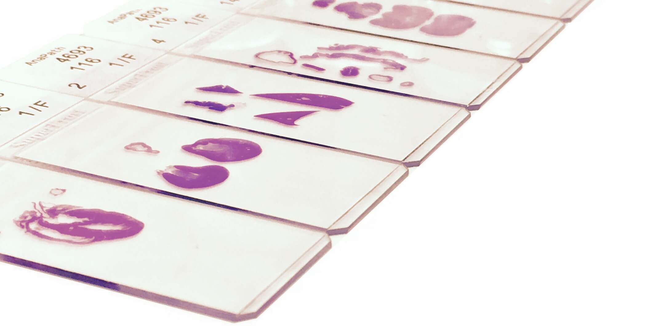
In anatomic pathology (anapath), slides refer to thin sections of tissue specimens that are mounted on glass slides for microscopic examination. These slides are prepared through a series of steps to ensure optimal visualization of cellular structures and tissue architecture.

Here's a general description of the process and the components of an anapath slide:
Specimen Collection:
Tissue samples are obtained from surgical procedures, biopsies, or autopsies. These samples may come from various organs or tissues of the body, depending on the clinical indication.
Fixation:
The tissue samples are placed in a fixative solution, such as formalin, to preserve cellular structures and prevent decay. Fixation helps to maintain the integrity of the tissue and prevents autolysis (self-digestion of tissues).
Processing:
After fixation, the tissue samples undergo a series of processing steps, including dehydration, clearing, and embedding in a paraffin wax or resin block. This process removes water from the tissue and replaces it with a medium that can be easily sectioned.
Microtomy:
The embedded tissue block is cut into thin sections (usually 3-5 micrometers thick) using a microtome. These sections are mounted onto glass slides, typically coated with an adhesive substance like albumin or poly-L-lysine, to ensure tissue adhesion.
Staining:
The mounted tissue sections are stained using various histological stains to enhance contrast and highlight specific cellular structures. Hematoxylin and eosin (H&E) stain is the most commonly used stain in anapath, which stains cell nuclei blue-purple (hematoxylin) and cytoplasm and extracellular structures pink (eosin). Other special stains may be used to highlight particular tissue components or pathological features.
Cover Slipping:
Once stained, the slides are coverslipped by placing a thin glass coverslip over the tissue section. This protects the specimen and helps to minimize distortion during microscopic examination.
Microscopic Examination:
The prepared slides are examined under a light microscope by pathologists or trained laboratory professionals. They analyze the tissue morphology, cellular architecture, and any abnormalities or pathological changes present.
Overall, anapath slides are essential diagnostic tools in pathology, providing detailed information about tissue structure and aiding in the diagnosis and management of various diseases and conditions.

The OxiSelect™ Comet Assay Slide is a specialized tool used in the comet assay, also known as single-cell gel electrophoresis. This technique is widely employed to assess DNA damage in individual cells. Here's an overview of the OxiSelect™ Comet Assay Slide:
Purpose:
The comet assay is a sensitive method for quantifying DNA damage and repair at the level of individual cells. It's particularly valuable in research related to genotoxicity, DNA repair mechanisms, and evaluating the effects of various agents on DNA integrity.
Design:
The OxiSelect™ Comet Assay Slide is designed to accommodate the comet assay in a single-slide format. It typically consists of a transparent substrate compatible with fluorescence microscopy, allowing for the visualization of stained DNA within individual cells.
Material:
The slide material is optimized to minimize background fluorescence and ensure accurate detection of comet tails, which represent DNA damage. It may be coated with a thin layer of agarose gel, providing a supportive matrix for embedding cells and facilitating electrophoresis.
Usage:
To perform the comet assay using the OxiSelect™ Comet Assay Slide, cells are embedded in agarose gel on the slide, lysed to remove cellular membranes, subjected to electrophoresis to separate fragmented DNA, and stained with a fluorescent dye such as ethidium bromide or SYBR Green. Under fluorescence microscopy, intact DNA appears as a compact nuclear region, while damaged DNA migrates away from the nucleus, forming a comet-like tail.
Analysis:
Image analysis software is often used to quantify DNA damage by measuring parameters such as comet tail length, tail intensity, and percentage of DNA in the tail. These measurements provide quantitative data on the extent of DNA damage in individual cells and can be used to compare experimental conditions or treatments.
There are no products listed under this category.
